

SABITA Research Highlights | June 2026
Recent publications from SABITA researchers highlight advances across neuroscience, biomedical science, and translational research.
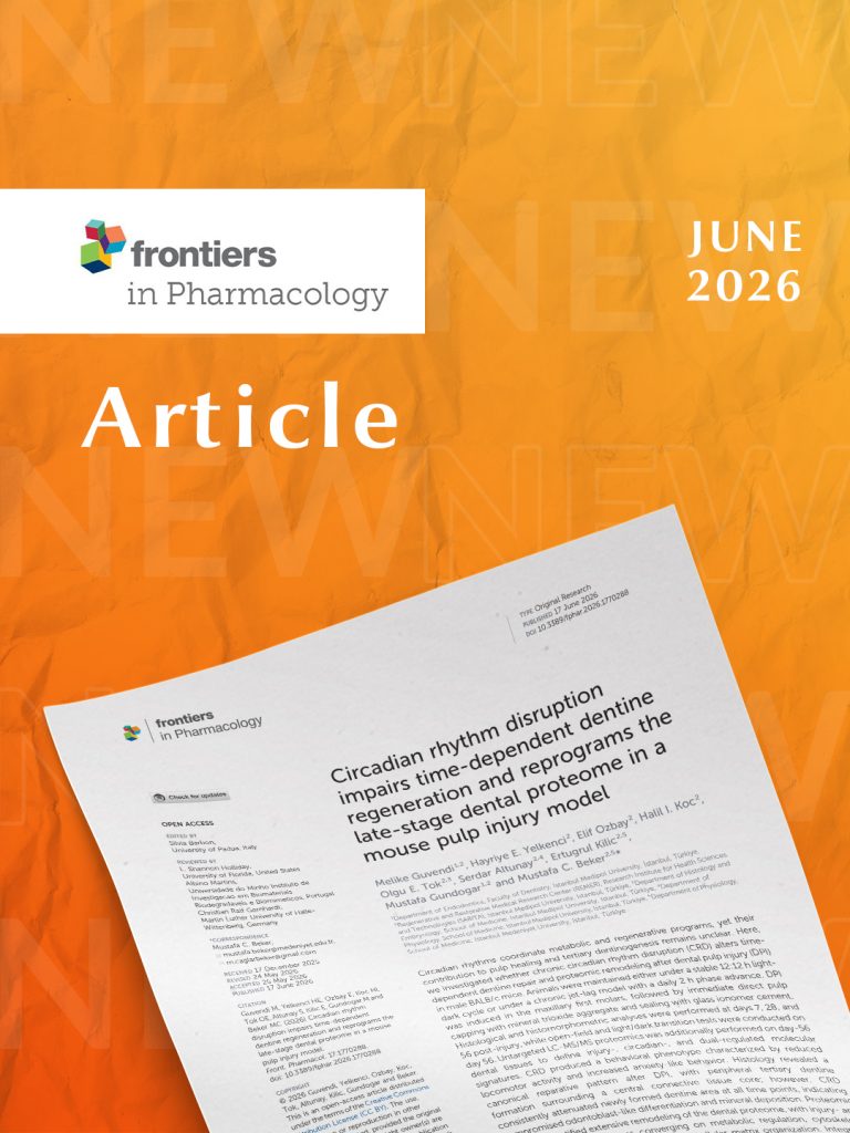
Circadian rhythm disruption impairs time-dependent dentine regeneration and reprograms the late-stage dental proteome in a mouse pulp injury model
Could your body clock influence how teeth heal?
Circadian rhythms regulate many physiological processes, from metabolism to tissue repair. But could disruptions to our biological clock also affect how dental tissues regenerate after injury?
A new study involving researchers from SABITA demonstrates that chronic circadian rhythm disruption impairs dentine regeneration and reshapes the molecular landscape of dental repair in a mouse model of pulp injury. Using a chronic jet-lag model, researchers evaluated how circadian disruption influenced healing after dental pulp injury treated with vital pulp therapy. Histological analyses showed that mice exposed to disrupted circadian rhythms consistently formed less tertiary dentine than animals maintained under a normal light–dark cycle, indicating reduced regenerative capacity over time. To better understand the underlying biology, the team performed untargeted LC-MS/MS proteomic analysis of dental tissues. The results revealed extensive molecular remodeling involving metabolic pathways, cytoskeletal organization, immune responses, and extracellular matrix regulation. Integrated analyses identified 204 injury-specific proteins, 128 circadian-specific proteins, and 86 proteins regulated by both injury and circadian disruption, highlighting a molecular intersection between tissue regeneration and biological timing.
Importantly, only a subset of these shared proteins was directly altered when circadian disruption occurred during tissue repair, suggesting that circadian misalignment selectively reprograms regenerative processes rather than globally altering the injury response.
Overall, the findings suggest that circadian rhythm integrity is an important biological factor influencing pulp healing and dentine regeneration. While further clinical studies are needed to determine whether similar effects occur in humans, this work provides new insight into how chronobiology may contribute to future regenerative endodontic strategies.
SABITA Researchers: Melike Güvendi, Hayriye E. Yelkenci, Elif Ozbay, Halil İbrahim Koç, Olgu E. Tok, Mustafa Gündoğar, Mustafa C. Beker
*This study was conducted in collaboration with researchers from international partner institutions. Full author list available in the original publication.
https://www.frontiersin.org/journals/pharmacology/articles/10.3389/fphar.2026.1770288/full
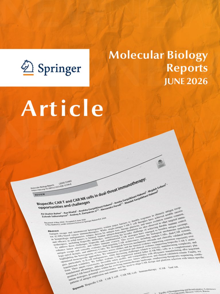
Bispecific CAR-T and CAR-NK cells in dual-threat immunotherapy: opportunities and challenges
Can dual-targeting CAR therapies overcome cancer immune escape?
Chimeric antigen receptor (CAR)-based immunotherapies have transformed the treatment of several blood cancers. However, long-term success is often limited by antigen escape—a process in which cancer cells lose or alter the target molecules recognized by engineered immune cells, allowing tumors to evade immune attack.
A new review involving researchers from SABITA, examines emerging bispecific CAR-T and CAR-NK cell strategies designed to address this challenge by targeting multiple tumor antigens simultaneously. Rather than focusing on a single therapeutic approach, the review provides a comprehensive evaluation of current evidence, biological rationale, and remaining translational challenges.
The authors discuss several next-generation CAR designs, including tandem CARs, dual-CAR systems, logic-gated CARs, and inhibitory CARs, each developed to broaden antigen recognition, improve tumor selectivity, and reduce immune evasion. The review emphasizes that although many dual-targeting strategies have demonstrated encouraging preclinical and early clinical results, evidence supporting consistent superiority over optimized single-target CAR therapies remains limited across different cancer types.
The review also highlights the growing potential of CAR-engineered natural killer (CAR-NK) cells. Compared with CAR-T cells, CAR-NK cells offer several potential advantages, including innate antitumor activity, a favorable safety profile, and compatibility with allogeneic “off-the-shelf” manufacturing. At the same time, important challenges—including limited persistence, tumor infiltration, and the need for further clinical validation—remain to be addressed.
Rather than advocating a single universal solution, the authors argue that future success will likely depend on matching CAR design and cellular platform to tumor-specific patterns of antigen expression and immune escape, while integrating CAR therapies with combination and sequencing strategies within the broader immunotherapy landscape.
Overall, the review highlights both the opportunities and the remaining challenges in developing next-generation CAR-based immunotherapies capable of achieving more durable and personalized cancer treatment.
SABITA Researcher and Corresponding author: Mazdak Ganjalikhani Hakemi
*This study was conducted in collaboration with researchers from national partner institutions.
https://link.springer.com/article/10.1007/s11033-026-12144-8
Investigation of the effects of balance exercises on visuospatial skills using EEG brain oscillations
Can balance training reshape the brain’s visuospatial networks?
Balance training is widely recognized for improving postural control, but could different types of balance exercises also influence how the brain processes spatial information?
A new study involving researchers from SABITA, investigated how structured balance exercises and video-based balance games affect visuospatial attention and mental rotation in healthy young adults using electroencephalography (EEG).
Thirty healthy participants were randomly assigned to one of two six-week exercise programs, completing supervised sessions twice a week. Before and after the intervention, participants performed visuospatial attention and mental rotation tasks while their brain activity was recorded using EEG to examine changes in neural oscillations associated with cognitive processing.
The researchers found that both exercise programs influenced visuospatial processing, but through different patterns of brain activity. During the visuospatial attention task, video-based balance training was associated with reduced occipital theta activity, whereas structured balance exercises showed an increase in the same neural oscillations. During the mental rotation task, the two interventions also produced distinct changes in high-alpha oscillations within centroparietal brain regions.
Behaviorally, participants in both groups improved their mental rotation performance after the intervention, while EEG findings suggested that each training approach may engage different neurophysiological mechanisms supporting visuospatial cognition.
The authors emphasize that these findings suggest different forms of balance training may modulate visuospatial cognitive processes in distinct ways rather than indicating that one approach is universally superior. They also note that future studies including larger cohorts, longer interventions, and additional behavioral measures will be important to further clarify these mechanisms.
SABITA Researchers: Bahar GÜNTEKİN
*This study was conducted in collaboration with researchers from national partner institutions.
Heart-Specific Spinal and Vagal Afferents: Transcriptomic Signatures and Optogenetically Modulated Functional Coupling With Cardiomyocytes
How does the heart communicate with the nervous system?
The heart continuously sends sensory information to the brain, helping regulate cardiovascular function. Yet, the molecular identity of the sensory neurons responsible for this communication has remained largely unknown.
A new study by researchers from SABITA, provides one of the most comprehensive characterizations to date of heart-specific sensory neurons, combining transcriptomics with functional analyses to investigate how these neurons interact with heart muscle cells.
Using retrograde labeling and fluorescence-activated cell sorting (FACS), the researchers isolated sensory neurons that specifically innervate the heart from both the dorsal root ganglion (DRG) and the nodose ganglion (NG). RNA sequencing revealed that these two neuronal populations possess distinct transcriptomic signatures, including differences in ion channels and G protein-coupled receptors that may reflect specialized physiological functions.
To investigate whether these neurons actively communicate with the heart, the team established neuron–cardiomyocyte co-cultures and combined calcium imaging with optogenetic stimulation. The experiments demonstrated functional interactions between heart-specific sensory neurons and cardiomyocytes, indicating dynamic neuro-cardiac communication rather than passive anatomical connections.
Together, the findings provide new insight into the molecular organization of cardiac sensory pathways and establish an experimental platform for studying how the nervous system monitors heart function. The authors suggest that these molecular signatures may support future investigations into cardiovascular reflexes, cardiac pain, and neuro-cardiac signaling mechanisms.
SABITA Researchers: T. Akgül Çağlar, Y. E. Kazci, Z. B. Durdu, S. Sahoglu Goktas, S. Bay, Emre Vatandaşlar, Esra Çagavi
Corresponding Author: Esra Çagavi
*This study was conducted in collaboration with researchers from national partner institutions.
Targeting Oral Tissue Regeneration With Standardized Centella asiatica Extract: Experimental Insights From Human Fibroblast Cultures
Can a medicinal plant support future oral tissue regeneration?
Natural compounds are increasingly being investigated for their potential use in regenerative medicine. Before such materials can be evaluated for therapeutic applications, their safety and compatibility with human cells must first be established.
Researchers from SABITA, evaluated the biocompatibility of a standardized Centella asiatica extract using cultured human dermal fibroblasts, a widely used in vitro model for connective tissue research.
The team exposed fibroblast cultures to six different concentrations of the extract, ranging from 5 to 200 µg/mL, and assessed cell viability using the MTT assay. Untreated cells served as the negative control, while Triton X-100 was used as a positive cytotoxicity control.
Across all tested concentrations, the extract maintained high levels of cell viability, with no statistically significant reduction compared with untreated cells. Furthermore, nonlinear regression analysis revealed no dose-dependent cytotoxic effect, indicating that increasing concentrations of the extract did not adversely affect fibroblast survival.
These findings suggest that the standardized Centella asiatica extract is biocompatible with healthy human fibroblasts under the experimental conditions tested. While additional preclinical and clinical studies are needed to evaluate its regenerative potential, the results provide an important foundation for future research on oral soft tissue regeneration and wound healing.
SABITA Researcher: Zeynep Budak
*This study was conducted in collaboration with researchers from national and international partner institutions. Full author list available in the original publication.
Oleuropein as a Potential Enhancer of Maxillofacial Flap Healing: In Vitro Evidence From Fibroblast Cultures
Could a natural olive compound support future regenerative therapies?
Successful healing following maxillofacial surgery depends on healthy cellular responses that support tissue repair and wound integration. Before naturally derived compounds can be explored for regenerative applications, their compatibility with human cells must first be established.
Researchers from SABITA, investigated the biocompatibility of oleuropein, a naturally occurring phenolic compound found in olive leaves and olives, using cultured human dermal fibroblasts.
Human fibroblasts were exposed to oleuropein at concentrations ranging from 10 to 200 µM, and cell viability was evaluated using the MTT assay. Untreated cells served as the negative control, while Triton X-100 was included as a positive cytotoxicity control to verify assay performance.
Across all tested concentrations, oleuropein maintained fibroblast viability without causing statistically significant reductions in cellular metabolic activity compared with untreated cells. In addition, dose-response analysis revealed no concentration-dependent cytotoxicity, indicating that increasing oleuropein concentrations did not adversely affect cell survival.
These findings suggest that oleuropein is biocompatible with human dermal fibroblasts under the experimental conditions examined. While further preclinical and clinical studies are required to investigate its effects on tissue repair and maxillofacial flap healing, the study provides important evidence supporting its cellular safety for future regenerative medicine research.
SABITA Researchers: Zeynep BUDAK
*This study was conducted within SABITA and Istanbul Medipol University research units.
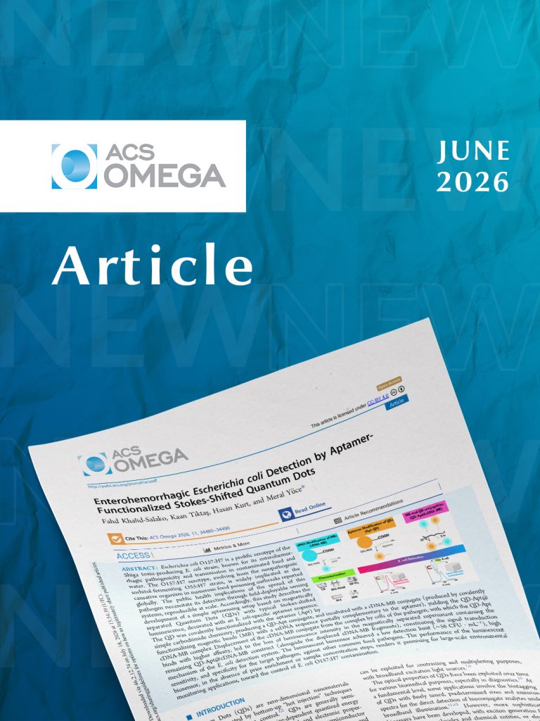
Enterohemorrhagic Escherichia coli Detection by Aptamer-Functionalized Stokes-Shifted Quantum Dots
Can foodborne bacteria be detected in under an hour?
Foodborne pathogens continue to pose a major public health challenge worldwide, highlighting the need for rapid, accurate, and field-deployable detection technologies. Conventional laboratory methods often require specialized equipment, trained personnel, and lengthy processing times, limiting their use in on-site monitoring.
Researchers including Hasan Kurt from SABITA, have developed a luminescent biosensor that combines quantum dots, DNA aptamers, and magnetic beads for the detection of Escherichia coli O157:H7, one of the most important foodborne bacterial pathogens.
The sensing platform uses quantum dots functionalized with an E. coli-specific DNA aptamer. When the target bacteria bind to the aptamer, a complementary DNA–magnetic bead complex is displaced, leading to a measurable decrease in fluorescence intensity. This optical signal provides the basis for detecting the presence of the pathogen.
Laboratory testing demonstrated that the biosensor detected E. coli O157:H7 with a limit of detection of approximately 56 colony-forming units (CFU) per milliliter, while maintaining high specificity against several other common foodborne bacteria, including Staphylococcus aureus, Salmonella typhimurium, Listeria monocytogenes, and Pseudomonas aeruginosa. Notably, the assay achieved this performance without prior enrichment or sample concentration, and the complete detection process required approximately 45 minutes.
The authors suggest that the combination of quantum dots and aptamers offers advantages over conventional antibody-based sensing approaches, including improved stability and reproducibility. They also note that the platform may provide a foundation for future multiplex biosensors capable of detecting multiple foodborne pathogens simultaneously by using quantum dots with different emission wavelengths.
Although the current study was validated primarily under laboratory buffer conditions, the findings support the potential of this biosensing strategy for future environmental monitoring and food safety applications. Further studies in complex food matrices and continued miniaturization will be important steps toward practical deployment.
SABITA Researcher : Hasan Kurt
*This study was conducted in collaboration with researchers from international partner institutions. Full author list available in the original publication.
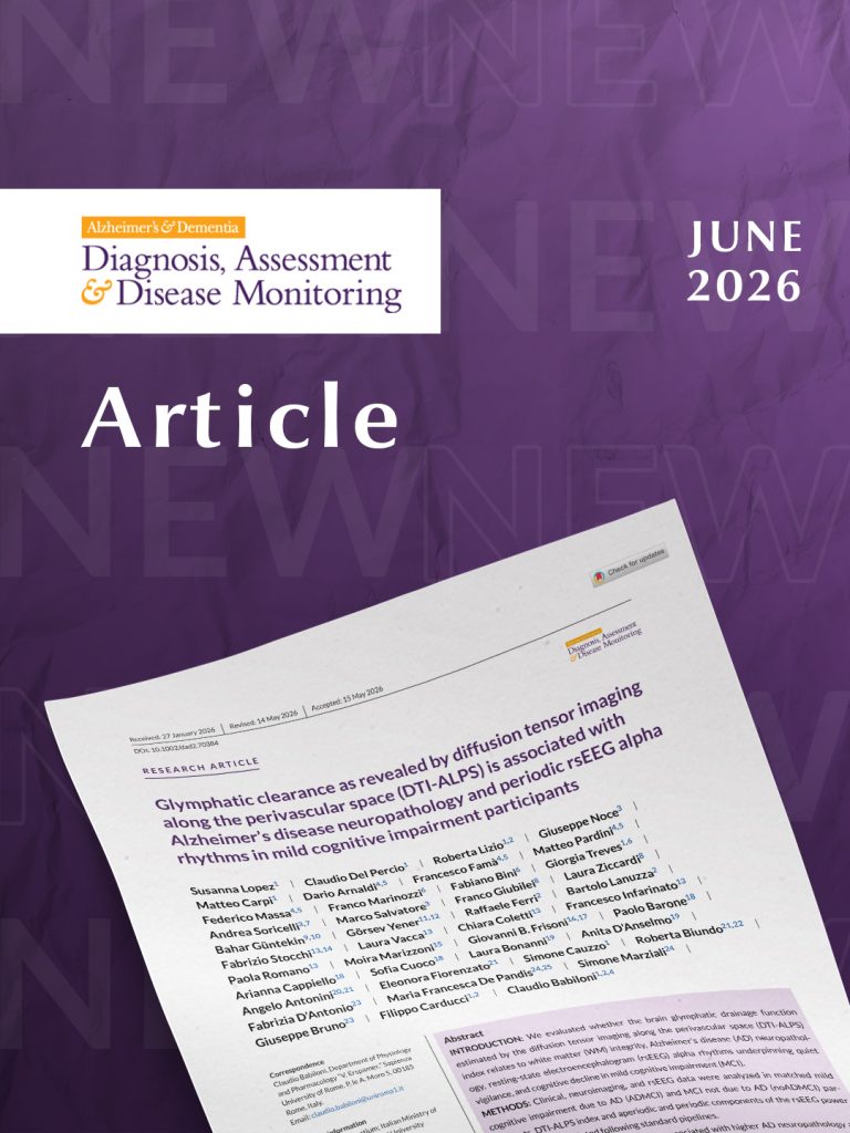
Glymphatic clearance as revealed by diffusion tensor imaging along the perivascular space (DTI-ALPS) is associated with Alzheimer’s disease neuropathology and periodic rsEEG alpha rhythms in mild cognitive impairment participants
Could the brain’s waste clearance system help explain early Alzheimer’s disease?
The brain possesses a specialized waste clearance network known as the glymphatic system, which helps remove metabolic waste products, including proteins associated with Alzheimer’s disease. Growing evidence suggests that disruption of this system may contribute to neurodegeneration, but its relationship with brain function during the early stages of Alzheimer’s disease remains under investigation.
Researchers, including Prof. Dr. Bahar Güntekin from SABITA, investigated whether impaired glymphatic function is associated with Alzheimer’s disease pathology, brain electrical activity, and cognitive performance in individuals with mild cognitive impairment (MCI). The study analyzed clinical, MRI, diffusion tensor imaging (DTI), cerebrospinal fluid biomarkers, and resting-state EEG data from participants with MCI due to Alzheimer’s disease and non-Alzheimer’s MCI.
To estimate glymphatic function, the researchers used Diffusion Tensor Imaging Along the Perivascular Space (DTI-ALPS), a non-invasive MRI-based imaging approach proposed as an indirect marker of glymphatic clearance. Participants with prodromal Alzheimer’s disease showed lower DTI-ALPS values, suggesting reduced glymphatic activity compared with individuals whose cognitive impairment was not attributed to Alzheimer’s disease.
Across the study population, lower DTI-ALPS values were associated with greater white matter lesions, more pronounced Alzheimer’s disease neuropathology, reduced posterior alpha brain rhythms measured by EEG, and poorer memory performance. These findings suggest that impaired glymphatic function may be linked not only to molecular changes associated with Alzheimer’s disease but also to alterations in functional brain networks involved in attention, vigilance, and cognition.
The authors emphasize that DTI-ALPS does not directly measure glymphatic flow and should be considered an indirect imaging marker. Because the study is cross-sectional, it cannot determine whether impaired glymphatic clearance causes cognitive decline or reflects ongoing neurodegenerative processes. Longitudinal studies will be needed to clarify its prognostic value and potential role in disease progression.
The findings contribute to the growing body of research exploring how vascular health, waste clearance mechanisms, and neural network function interact during the earliest stages of Alzheimer’s disease, potentially supporting future biomarker development and earlier disease monitoring.
SABITA Researchers: Bahar GÜNTEKİN
*This study was conducted in collaboration with researchers from national and international partner institutions. Full author list available in the original publication.
https://alzjournals.onlinelibrary.wiley.com/doi/10.1002/dad2.70384
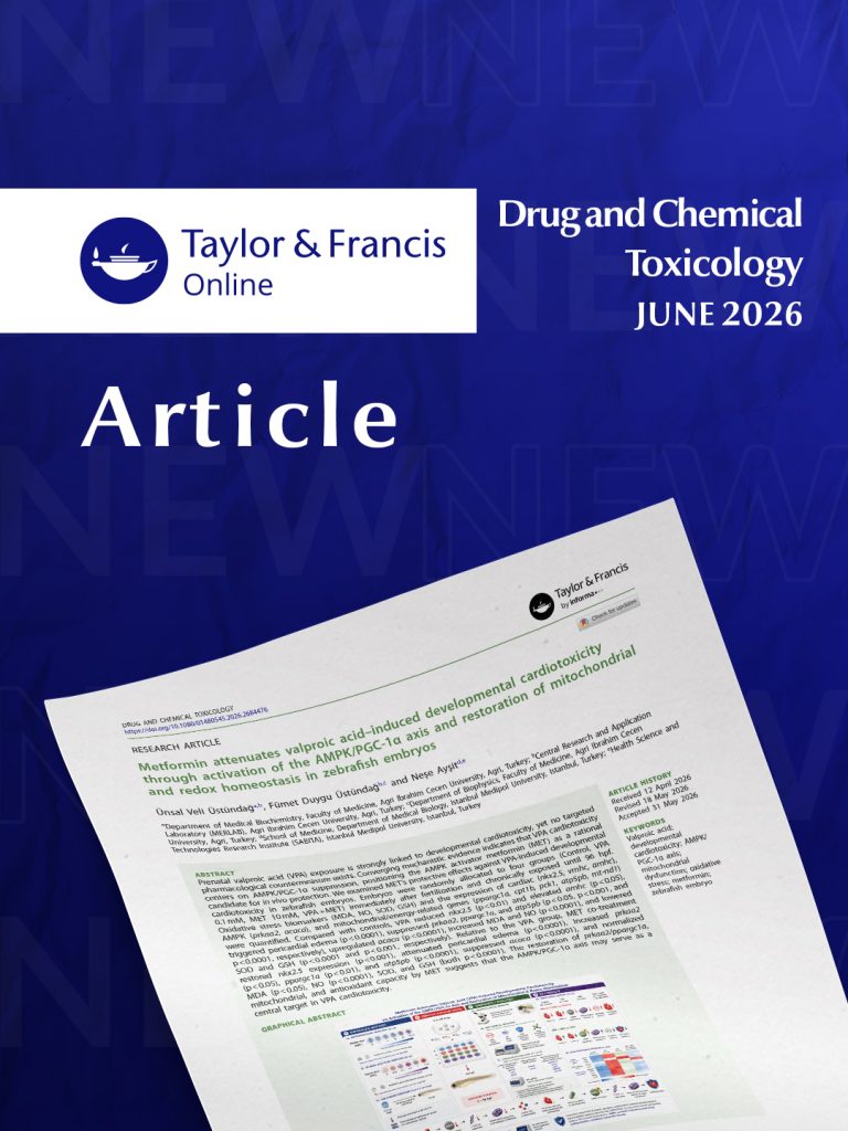
Metformin attenuates valproic acid-induced developmental cardiotoxicity through activation of the AMPK/PGC-1α axis and restoration of mitochondrial and redox homeostasis in zebrafish embryos
Can a metabolic pathway help protect the developing heart?
Valproic acid (VPA) is an effective medication for epilepsy and several neurological disorders, but prenatal exposure has been associated with developmental abnormalities, including defects affecting the heart. Understanding the biological mechanisms underlying these effects may help identify new strategies for reducing developmental toxicity.
Researchers, investigated whether metformin, a widely used metabolic drug, could reduce VPA-induced developmental cardiotoxicity in zebrafish embryos, an established model for studying early vertebrate development.
Embryos were exposed to valproic acid, metformin, or a combination of both from fertilization until 96 hours post-fertilization. The researchers evaluated cardiac development, oxidative stress, and the expression of genes involved in energy metabolism, mitochondrial function, and cardiac development.
Compared with embryos exposed to VPA alone, those receiving metformin co-treatment showed reduced pericardial edema and restoration of key cardiac developmental markers, including nkx2.5. Metformin also reversed changes in genes associated with the AMPK/PGC-1α signaling pathway, improved the expression of mitochondrial energy metabolism genes, and reduced oxidative stress by normalizing antioxidant and redox biomarkers.
These findings suggest that activation of the AMPK/PGC-1α pathway may help preserve mitochondrial function and cellular redox balance during VPA-induced developmental stress. The results provide new mechanistic insight into how metabolic pathways may influence embryonic heart development under toxic conditions.
The authors emphasize that these findings were obtained in zebrafish embryos, and further studies in mammalian models and clinical settings will be necessary before any therapeutic implications for human pregnancy can be considered.
SABITA Researchers: Neşe AYŞİT
*This study was conducted in collaboration with researchers from national partner institutions. Full author list available in the original publication.
https://www.tandfonline.com/doi/full/10.1080/01480545.2026.2684476
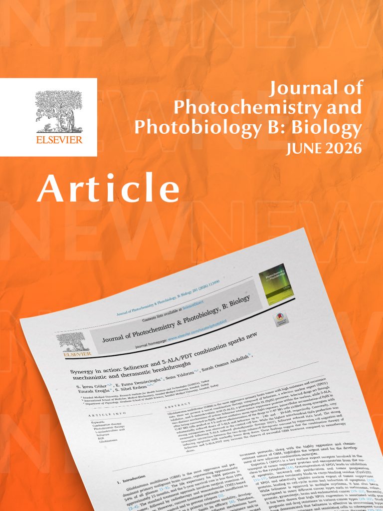
Synergy in action: Selinexor and 5-ALA/PDT combination sparks new mechanistic and theranostic breakthroughs
Can two complementary therapies work better together against glioblastoma?
Glioblastoma (GBM) is the most aggressive primary brain tumor, characterized by rapid progression, treatment resistance, and frequent recurrence. Despite advances in surgery, radiotherapy, and chemotherapy, long-term disease control remains a major clinical challenge.
Researchers from SABITA, investigated a novel combination strategy that pairs Selinexor, a selective inhibitor of nuclear export, with 5-aminolevulinic acid (5-ALA)-based photodynamic therapy (PDT). The approach was designed to simultaneously target complementary mechanisms involved in tumor survival and oxidative stress.
Using the human glioblastoma U-87 MG cell line, the researchers found that combining Selinexor with 5-ALA/PDT produced strong synergistic anti-tumor effects, allowing substantially lower doses of both agents to achieve therapeutic activity compared with either treatment alone. Interestingly, this synergistic interaction was not observed in the non-cancer control cell line, where the combination showed marked antagonism, suggesting greater selectivity toward tumor cells.
Mechanistic analyses revealed that the combination suppressed glioblastoma cell migration, promoted programmed cell death, and restored molecular pathways involved in mitochondrial function and cellular stress responses. While photodynamic therapy generated oxidative stress through mitochondrial reactive oxygen species, Selinexor appeared to modulate this response while enhancing overall anti-tumor activity through inhibition of the XPO1-mediated nuclear export pathway.
The findings suggest that combining nuclear export inhibition with photodynamic therapy may represent a promising strategy for enhancing therapeutic responses against glioblastoma while reducing the doses required for each treatment. However, the study was conducted in cultured cancer cells, and further validation in animal models and clinical studies will be required before this approach can be considered for patient care.
SABITA Researcher: S. İrem Güler, E. Esma Demircioglu, Sena Yıldırım, Sarah Osama Abdallah, Emrah Eroğlu, S. Sibel Erdem
*This study was conducted in collaboration with researchers from international partner institutions. Full author list available in the original publication.
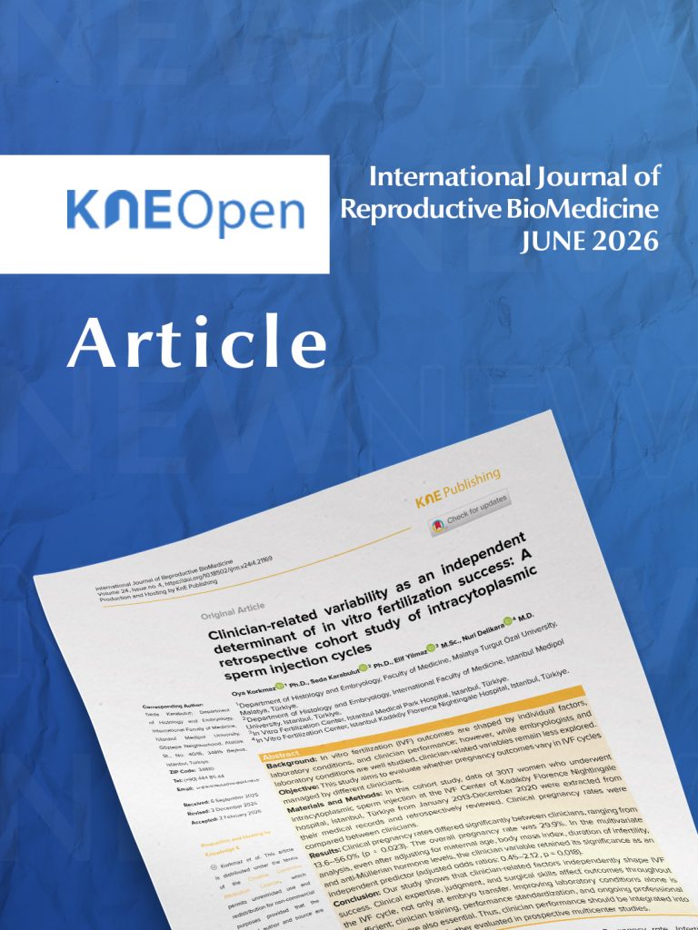
Clinician-related variability as an independent determinant of in vitro fertilization success: A retrospective cohort study of intracytoplasmic sperm injection cyclesClinician-related variability as an independent determinant of in vitro fertilization success: A retrospective cohort study of intracytoplasmic sperm injection cycles
Beyond the laboratory: Does clinician performance influence IVF success?
In vitro fertilization (IVF) outcomes are influenced by many factors, including patient characteristics, laboratory procedures, and embryo quality. While laboratory performance has been extensively studied, the contribution of clinician-related factors has received comparatively less attention. A new study suggests that clinical expertise may also play an important role in treatment success.
Researchers from Istanbul Medipol University and collaborating fertility centers analyzed 3,017 intracytoplasmic sperm injection (ICSI) cycles performed between 2013 and 2020 to investigate whether pregnancy outcomes differed among clinicians working within the same IVF center. By evaluating treatments performed under comparable laboratory conditions, the study aimed to isolate the contribution of clinician-related factors.
Clinical pregnancy rates varied significantly among the 22 clinicians included in the analysis, ranging from 13.6% to 56.0%, with an overall pregnancy rate of 29.9%. Importantly, these differences remained statistically significant even after adjusting for maternal age, body mass index, infertility duration, and anti-Müllerian hormone (AMH) levels, indicating that clinician-related factors independently contributed to treatment outcomes.
The authors suggest that IVF success depends on a series of clinical decisions extending beyond embryo transfer. Individualized ovarian stimulation protocols, medication adjustments, oocyte retrieval techniques, procedural skills, and coordination with embryology teams may all influence the likelihood of achieving pregnancy.
Rather than attributing success to any single procedure, the findings highlight the importance of consistent clinical practice, structured training, and quality assurance throughout the entire IVF process. The authors propose that clinician-related performance metrics could complement existing laboratory quality indicators to support continuous improvement in assisted reproductive medicine.
Because this was a retrospective single-center study, the findings should be interpreted with appropriate caution. The study evaluated clinical pregnancy rather than live birth, and additional multicenter prospective studies will be necessary to confirm these observations and identify which aspects of clinical practice most strongly influence IVF outcomes.
SABITA Researcher: Seda Karabulut
*This study was conducted in collaboration with researchers from national and international partner institutions. Full author list available in the original publication.
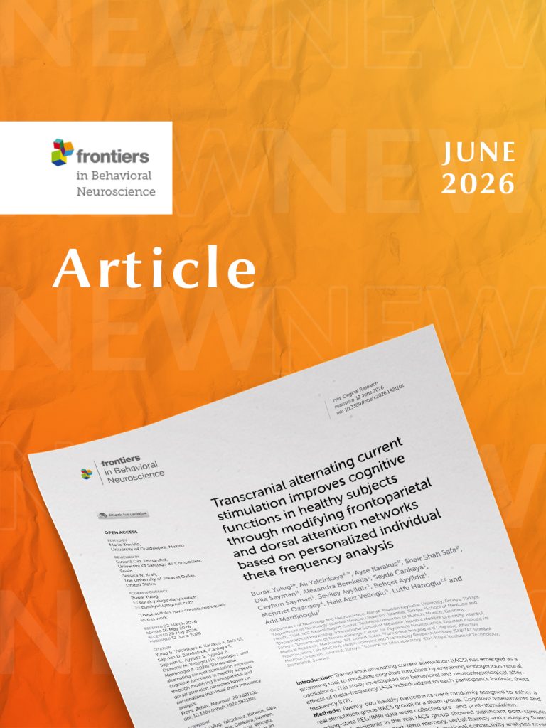
Transcranial alternating current stimulation improves cognitive functions in healthy subjects through modifying frontoparietal and dorsal attention networks based on personalized individual theta frequency analysis
Can non-invasive brain stimulation strengthen cognitive functions by aligning with the brain’s natural rhythms?
A new study by researchers from Istanbul Medipol University, SABITA, and collaborating institutions investigated whether personalized transcranial alternating current stimulation (tACS) could improve cognitive performance in healthy adults by targeting each individual’s natural theta brainwave frequency.
The randomized controlled study included 22 healthy participants, who received either active or sham stimulation over five consecutive days. Before and after the intervention, participants completed comprehensive neuropsychological assessments while undergoing electroencephalography (EEG) and resting-state functional MRI (fMRI) to examine changes in brain activity and functional connectivity.
Participants receiving personalized tACS demonstrated significant improvements in short-term memory, verbal fluency, and category fluency compared with the sham group. Brain imaging analyses also revealed changes in the frontoparietal and dorsal attention networks, two large-scale neural networks involved in executive function, attention, and memory processing.
Rather than simply measuring behavioral performance, the study combined EEG-informed stimulation with functional MRI to investigate how cognitive improvements may relate to changes in brain network organization. The findings suggest that individualized brain stimulation may influence functional connectivity in networks associated with higher cognitive functions.
The authors emphasize that these findings were obtained in a small group of healthy young adults and should be interpreted cautiously. Larger studies will be needed to determine whether similar approaches can benefit individuals with neurological disorders or cognitive impairment and to establish the long-term effects of personalized brain stimulation.