


Cagavi Lab/Frameworks
Disease Models of Congenital Arrhythmia
Cardiac disorders are the leading cause of death worldwide. Arrhythmia is the most common manifestation of cardiac disorders that can lead to life-threatening fibrillations. Hereditary forms of arrhythmia are classified under channelopathies and caused by ion channel mutations. We are interested in modeling congenital cardiac arrhythmia, particularly Long QT Syndrome subtypes, by generating patient-derived human induced pluripotent stem cells and differentiate them into cardiomyocytes (hiPSC-CMs). As a routine application in our lab, we reprogram mononucleated blood cells from healthy individuals and patients into iPSCs. Derivation of cardiac cultures from hiPSCs is carried out by manipulation of signal pathways by small molecules. We obtain hiPSC-CMs in high efficiency (below), analyze by morphological, molecular, and electrophysiological assays.
Systems Biology: Cardiac Nervous System Interactions
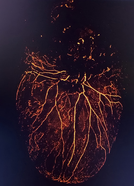
The heart is highly innervated with the autonomic nervous system to regulate its vital functions. To maintain cardiac homeostasis and for temporal regulation, there is active neural communication between the heart and the brain. Even though the motor control over the myocardium is well-characterized, the identity of cardiac sensory neurons and the extent of the information reported to the brain via these neurons are poorly understood. Information regarding the mechanical load, oxidative stress, chemical cues, and other signals of steady or diseased-state of the cardiac tissue are actively relayed to the central nervous system. Our current knowledge about spatial innervations of the heart is limited and functional characteristics of the neural fibers of different anatomic origins remain to be elucidated. To spatially and functionally to identify cardiac afferent neuronal networks and to investigate the mechanisms governing the afferent signal transmission, we are employing an intersectional approach using retrograde labeling strategies, transcriptome analysis, viral vectors, and transgenic mice models. Along with neuronal mapping of cardiac afferents, we utilize optogenetics and chemogenetics tools to drive specific activation of vagal and spinal afferents and investigate their respective impacts on electrophysiological activities of the heart.
In the case of cardiac disorders, neuronal innervations and accompanying cardiac-nervous system interactions undergo abnormal remodeling, contributing to cardiac pathophysiology. The majority of cardiac arrhythmia has been suggested to be related to the disorders in cardiac innervations. Nevertheless, there is still so much to learn about the physiology and molecular mechanisms behind the defects. Recently, iPSC based microfluidic platforms have gained attention due to their unique properties and advantages. In our lab, in addition to the above mentioned in vivo approaches, we are investigating the nature of cardiac-nervous system interactions in vitro and developed a modeling strategy with human iPSC-derived mature cells and microfluidic platforms to replicate the in vivo microenvironment.
Mitochondrial Bioenergy Metabolism in Diseased State
The bioenergy metabolism can be evaluated by recording glycolysis and aerobic respiration parameters. These analyses have the potential to reveal new insights into disease pathology and progress. We are currently developing tools and using Seahorse XF analyzer to measure oxygen consumption and extracellular acidification rates of iPSC derived mature cells to compare in vitro mitochondrial function.
Cardiac Immunology
Macrophages are a group of phagocytic cells residing in many organs. Interesting recent findings indicated unconventional roles for cardiac resident macrophages in regulating electrophysiological properties of cardiomyocytes in experimental animal models. We are currently generating human macrophages from monocytes by growth factor stimulation to evaluate their role in cardiac physiology in vitro.