


Advanced Microscopy
Advanced Microscopy Facility provides a well-equipped infrastructure to conduct advanced scientific research requiring a wide range of techniques from fluorescent imaging to nanometre-scale high-resolution visualisation technologies.
These microscopy techniques enable real-time imaging in tissue samples and cell cultures, as well as to produce detailed three-dimensional images of soft tissue and bone structure of live animals. The unparalleled sensitivity of the systems makes it the ideal live-cell imaging, allowing for high-speed acquisition, supporting both in vivo and in vitro studies.
The unit is equipped with confocal and multi-photon fluorescent microscopes, transmission and scanning electron microscopes, fluorescent stereomicroscope, microdissection microscopes and lightsheet microscope, as well as sample preparation techniques such as microtome, ultra-microtome, and frozen sectioning.
Advanced Microscopy Facility, with its wide range state-of-the-art medical devices, makes it easier for the researchers to conduct deep and dynamic research projects. Therefore, the process of developing novel methods, products or technologies from bench to bedside is supported with the technological amendments.
Equipment

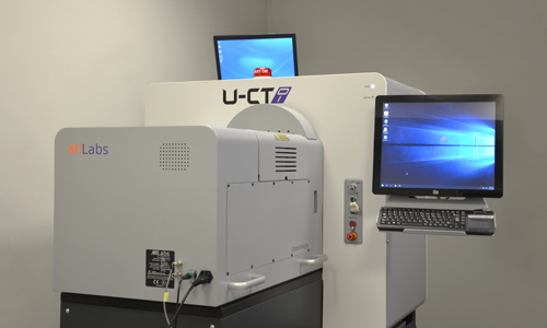
MILabs U-CT
Micro-Computed Tomography
X-ray-based 3D imaging enables researchers to investigate the three-dimensional microstructure of biological tissues and biomaterials.
At SABİTA, this technology supports research on bone microarchitecture, bone regeneration, biomaterial characterization, and tissue engineering.
Measurements and Analysis
- Bone Microarchitecture
- Cortical Bone Analysis
- Bone Mineral Density
- 3D Morphometric Analysis
- Vascular Imaging
- Dynamic Contrast-Enhanced CT
- Soft Tissue Imaging
- Longitudinal Imaging
Research Publications
Laboratory
Laboratory Name: In Vivo Imaging – Micro CT
Laboratory Location: North Campus, C-B48
Technical Support

Zeiss LSM 880 Airyscan
Laser Scanning Confocal Microscopy
The ZEISS LSM 880 with Airyscan is a laser scanning confocal microscope designed for high-resolution fluorescence imaging. Its advanced Airyscan detector enhances image sensitivity and spatial resolution, enabling detailed visualization of cellular and subcellular structures.
At SABİTA, this system supports advanced fluorescence microscopy, three-dimensional imaging, and the investigation of biological structures and cellular processes.
Measurements and Analysis
- Confocal Fluorescence Imaging
Airyscan Super-Resolution Imaging
3D Optical Sectioning and Reconstruction
Z-Stack Imaging
Multichannel Fluorescence Imaging
Time-Lapse Imaging
Colocalization Analysis
High-Sensitivity Fluorescence Detection
Research Publications
Laboratory
Laboratory Name: Advanced Microscopy Laboratory-2
Laboratory Location: North Campus, C-B32
Technical Support

MILabs U-CT
Micro-Computed Tomography
X-ray-based 3D imaging enables researchers to investigate the three-dimensional microstructure of biological tissues and biomaterials.
At SABİTA, this technology supports research on bone microarchitecture, bone regeneration, biomaterial characterization, and tissue engineering.
Measurements and Analysis
- Bone Microarchitecture
- Cortical Bone Analysis
- Bone Mineral Density
- 3D Morphometric Analysis
- Vascular Imaging
- Dynamic Contrast-Enhanced CT
- Soft Tissue Imaging
- Longitudinal Imaging
Research Publications
Laboratory
Laboratory Name: In Vivo Imaging – Micro CT
Laboratory Location: North Campus, C-B48