


Advanced Microscopy
Advanced Microscopy Facility provides a well-equipped infrastructure to conduct advanced scientific research requiring a wide range of techniques from fluorescent imaging to nanometre-scale high-resolution visualisation technologies.
These microscopy techniques enable real-time imaging in tissue samples and cell cultures, as well as to produce detailed three-dimensional images of soft tissue and bone structure of live animals. The unparalleled sensitivity of the systems makes it the ideal live-cell imaging, allowing for high-speed acquisition, supporting both in vivo and in vitro studies.
The unit is equipped with confocal and multi-photon fluorescent microscopes, transmission and scanning electron microscopes, fluorescent stereomicroscope, microdissection microscopes and lightsheet microscope, as well as sample preparation techniques such as microtome, ultra-microtome, and frozen sectioning.
Advanced Microscopy Facility, with its wide range state-of-the-art medical devices, makes it easier for the researchers to conduct deep and dynamic research projects. Therefore, the process of developing novel methods, products or technologies from bench to bedside is supported with the technological amendments.
Techniques Applied
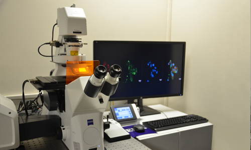
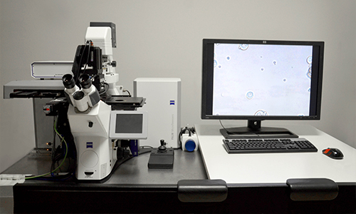
Confocal Microscopy
Confocal microscopy is an optical imaging technique for increasing optical resolution and contrast of a micrograph by means of using a spatial pinhole to block out-of-focus light in image formation. There are 3 different confocal microscopy systems according to their intended use.
Stereo Microscopy
Stereo Microscopy is a microscopic imaging technique used binocular microscope that gives a relatively low-power stereoscopic view of the subject. A method we enjoy using for rapid fluorescence imaging in our work.
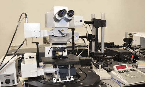
Multiphoton Microscopy
Multiphoton Microscopy is a fluorescence imaging technique that allows imaging of living tissue up to about one millimeter in thickness, with 0.64 μm lateral and 3.35 μm axial spatial resolution. A great method for high resolution imaging and manipulation in vivo and in vitro.
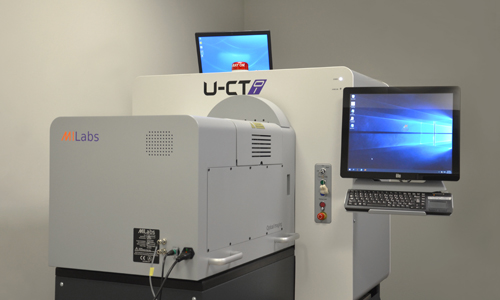
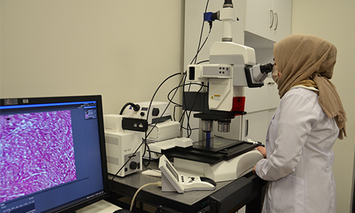
In Vivo Animal Imaging
In Vivo Animal Imaging is the visualization of living animals for research purposes. Micro-CT can have excellent spatial resolution, which can be up to 6 µm when combined with contrast agents.
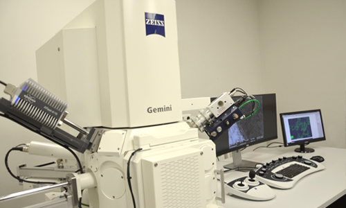
Electron Microscopy
Electron Microscopy is a technic that uses a beam of accelerated electrons as a source of illumination. As the wavelength of an electron can be up to 100.000 times shorter than that of visible light photons. Electron microscopy techniques that can perform both SEM and TEM imaging can be used in our fascility.
Microdissection
Microdissection is a method for isolating specific cells of interest from microscopic regions of tissue/cells/organisms At the same time, we can perform axotomy experiments on neurons with this technique.